
 Cryo-Electron Microscopy (Cryo-EM)
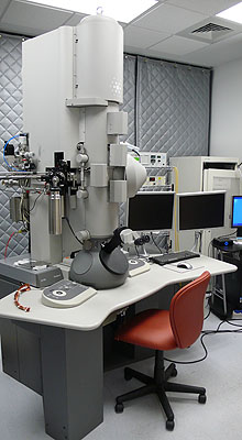
The cryo-EM suite at the Department of Structural Biology is located in the basement of BST3 and is furnished with state-of-the-art electron microscopes with full capability to carry out high resolution three-dimensional structural analysis of proteins, protein complexes, viruses, macromolecular assemblies, cellular organelles, and bacterial cells. At present, the cryo-EM facility comprises three Thermo Fisher (FEI) microscopes – a Titan Krios 3Gi equipped with a Falcon 3ec direct electron detector; a TF20 equipped with a TVIPS XF416 camera and Gatan cryoholders; and a T12 equipped with Gatan US 1000 and Orius CCD cameras. The Krios and TF20 are capable of low-dose “single-particle” and tomographic studies. The T12 Spirit may also be used for cryo-EM but is more usually used to examine room temperature specimens by negative stain EM. The facility is also equipped with a FEI Vitrobot for frozen-hydrated specimen preparation and other ancillary equipment for cryo-EM. A Bioquantum/K2 to extend the Krios, and an Aquilos cryoFIB mill, will be installed in the spring of 2020.
Conventional negative-stain electron microscopy provides two-dimensional (2D) projection images of the stain-embedded specimen in which the distribution of the heavy-metal stain represents the stain-excluding shape of the specimen. Cryo-EM offers a more direct representation of protein structure that avoids the dehydration and chemical modification of classical methods by immobilizing the specimen in a thin layer of vitrified (non-crystalline) buffer. By combining many images of identical objects imaged at different orientations, back-projection methods allow recovery of a model that represents the original three-dimensional object. Cryo-EM is proving to be an increasingly powerful tool by achieving atomic-resolution descriptions of protein complexes. Tomographic studies are also uniquely powerful in offering detailed structures of non-uniform objects such as organelles and offer insights into cellular and tissue organization that cannot otherwise be achieved. Cryo-EM bridges a critical gap in imaging in the biomedical size spectrum, the gap between molecules at atomic resolution by X-ray, NMR and now cryo-EM itself, and cells and tissues by light microscopic and conventional electron microscopic methods. This gap comprises a size range of considerable interest in biology and medicine that includes cellular protein machines, large protein assemblies and nucleic acid assemblies, small subcellular organelles and small bacteria. These objects are generally too large and/or too heterogeneous to be investigated by high resolution X-ray and NMR methods; yet the level of detail afforded by conventional light and electron microscopy is often not adequate to describe their structures at resolutions high enough to be useful in understanding the chemical basis of biological function.
Two areas in 3D cryo-electron microscopy are of particular note: “single particle” microscopy and electron tomography.
Single particle cryo-EM
Biological specimens are radiation sensitive, requiring very low-intensity images that have very noisy background. To overcome this problem, images recorded from thousands of identical copies of specimens randomly oriented relative to the electron beam are combined and averaged. This method is referred as “single particle” microscopy as the particles are “free-standing” or “single” as opposed to participating in a higher level of assembly such as a crystal. Many structures have been determined by cryo-EM, recently to atomic resolution, and covering sizes from 200kDa to 200MDa and higher. Most importantly, these structures allow interpretation of underlying tertiary, secondary and primary structural elements as well as interfaces between subunits, and usually in a native, functional context. This methodology can be used to determine the structures of complexes when they are too large and/or too heterogeneous to be investigated by high resolution X-ray and NMR methods. Structures that do not reach atomic resolution will nonetheless provide molecular envelopes for large complexes into which subunit atomic models determined either by X-ray crystallography, NMR or cryo-EM itself can be docked, yielding pseudo-atomic resolution models that are useful for understanding architecture and function.
Cryo-electron tomography
Electron tomography (ET) involves structure determination of “one-of-a-kind” objects such as whole cells and subcellular organelles, in which averaging methods are not applicable. The underlining principle is similar to computerized tomography (CT). Instead of rotating the radiation source and detector around the object, in ET, it is the specimen that is tilted under the incident electron beam and a series of projection images over a range of orientations are recorded and combined computationally to reconstruct the 3D structure of the object. A fundamental limitation to the studies of whole-cell specimens is that useful images can only be recorded from the thinnest regions of the cell (typically <0.4 µm) or from whole cells that are small and thin. Two approaches may be employed in cellular studies.
(i) ET of fixed and stained specimens at room temperature. The cells can be fixed (cryo or chemically) and embedded and sectioned. This has been a successful approach used to yield information on the membrane connectivity of cells and organelles or for recognition of cellular location of large and easily-stained objects.
(ii) Cryo-ET of unstained specimens rapidly vitrified at liquid nitrogen temperatures. This approach allows obtaining structural information about protein complexes and cellular assemblies at near native state of the cell.