 X-ray Crystallography
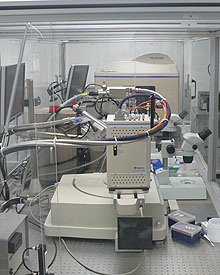
The 1500 square foot X-ray crystallography suite on the first floor of BST3 houses the main equipment and is supplemented by two environmentally controlled rooms to grow, store, and monitor crystals, at 18°C and at 4°C temperatures. One X-ray generator and two detectors are controlled from a central room to initiate data collection, while the control room also permits direct entry to and constant visualization/adjustment of experiments going on in the adjacent generator rooms. Rigaku X-ray equipment is provided for variety of experimental sample types. The high intensity, FR-E SuperBright generator and focusing optics provide a combination of existing rotating anode expertise and microfocus X-ray generation technology, yielding a flux within the same order of magnitude as a 2nd generation synchrotron bending magnet. Both ports on each generator are utilized and detectors include a Saturn 944 CCD, and a high throughput RAXIS HTC image plate. All ports are equipped with VariMax optics to provide high intensity and highly focused X-ray beams, as well as X-stream 2000 cryogenic systems. Ports supporting the CCD detectors utilize HF (high flux) optics with computerized slit control, while HR (high resolution) optics are provided on ports supporting the image plate detectors. This mix allows for high quality data collection on small and normal sized crystals, as well as on crystals with large unit cells typically containing very large proteins, viruses, or protein-protein assemblies. In-house liquid and dry nitrogen gas is supplied for all cryogenic systems, as well as helium for beam path purging.
In addition, the department is a member of a collaborative access team (SER-CAT, sector 22) at the APS (Advanced Photon Source) in Argonne, IL, to guarantee regular, 3rd generation synchrotron access to X-ray facility members when the absolute highest resolution possible is needed, only extremely small or very weakly diffracting crystals are available, or MAD/SAD phasing experiments are needed at non-standard wavelengths.
Within the main X-ray crystallography suite are located two additional labs, devoted to mounting, and computational analysis. The optical microscopy room, facilitates crystal examination/mounting/freezing and manual monitoring of crystallization set-ups. It contains multiple Olympus SZX12, SZX7, and SZ-61 microscopes, with high-end microscopes equipped with high-resolution cameras interfaced with LCD monitors and workstations for image capturing and recording. The computational room, contains multiple workstations, to facilitate rapid processing and analysis of incoming or previously collected data.
For Small-Angle X-ray Scattering studies, an Anton Paar SAXSess mc2 instrument with a sealed tube generator is also housed in the microscopy/mounting lab. This instrument includes a unit for controlling the temperature of the sample with Peltier heating/cooling, allowing for measurement at temperatures between -30°C and 120°C with a ± 0.1°C accuracy; a μ-cell and sample holder for the measurement of very small sample volumes; and a Mythen Detector for SAXSess, which unlike standard imaging plate detectors, allows for time-resolved and automated SAXS experiments.
Collectively, the X-ray crystallography facility and its resources provide everything needed to carry out state of the art crystallographic analysis of macromolecules. Professor William Furey serves as Co-Director of the X-ray facility, while Mr. Doowon Lee is the day-to-day facility manager dealing with maintenance, training, and user scheduling.